The patient may already know that when a mass is detected on an ultrasound the technician will then run a test to see if the mass has a vascular network since tumors do have their own vascular. Breast mris are usually reserved for women with an elevated risk of developing breast cancer have already had breast cancer or as a follow up study to inconclusive findings on a mammogram ultrasound and exam.

Targeted Breast Ultrasound Can Reduce Biopsies For Women Under Forty
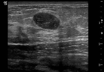
Ultrasound is a good tool for telling the difference between lumps that are fluid filled called cysts and those that are solid which can indicate cancer or another benign breast condition such as fibroadenoma.

Benign breast cancer on ultrasound. Breast ultrasound cancer vs benign this would all be very predictive of invasive ductal carcinoma and the lesion will require a biopsy for diagnosis. About 25 of all breast masses turn out to be cysts. They can be very tiny or they can be large enough to feel through the skin or see on an imaging test a grossly evident cyst or gross cyst.
The ultrasound can pick up a cyst in a breast. She wants the ultrasound for supplemental screening due to a genetically high risk of breast cancer. A variety of tests are used for the diagnosis of breast.
We aim to demonstrate the spectrum of us imaging findings in lactating. Ultrasound can tell the difference between solid masses that might be cancer versus fluid filled cysts which are generally not cancerous. On an mri findings that suggest breast cancer include a mass with spiculated margins rim enhancement around the mass or what is.
Most breast lesions diagnosed in these patients are benign but the diagnosis of breast cancer is a challenge. Radiographic features ultrasound malignant characteristics with positive predictive values. When breast problems occur patients are often referred for an ultrasound us scan.
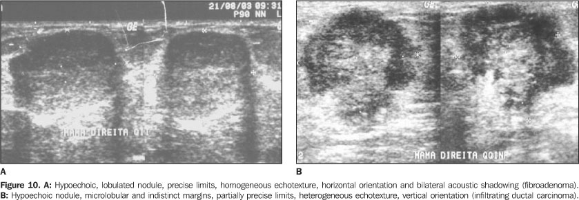
Benign and malignant characteristics of breast lesions at ultrasound allow the classification as either malignant intermediate or benign based on work published by stavros et al. Non cancerous and cancerous breast lumps can be very different from each other when it comes to how they feel during a breast exam and what they look like in imaging tests. Breast ultrasound can detect lumps that may not be seen on mammograms and they can also note changes in the breasts in women with dense breast tissue.
Structural changes during lactation make breast physical examination difficult. However a number of benign breast changes mimic breast cancer so it sometimes takes further testing to know for sure whats going on in your breast. Ultrasound is now used to evaluate masses seen on mammography and magnetic resonance imaging mri and may also be used to evaluate clinical breast symptoms such as palpable masses focal.
Most breast cysts are benign and do not increase your risk of breast cancer. Breast cysts are round or oval structures filled with fluid. However with major advances in ultrasound technology during the past 20 years ultrasound can also now distinguish benign and malignant solid breast masses.
Speculations corner margin hypoechoic higher than the width of the speculations breast tumors often represent tentacles or desmoplastic reaction.

Seeking To Reduce Biopsies For Breast Tumors Through Novel Imaging Technology Mckelvey School Of Engineering At Washington University In St Louis

Mammogram Images Normal Abnormal And Breast Cancer

Breast Ultrasound Atlanta Breast Screening

Radiologia Brasileira Ultra Sonografia Mamaria Avaliacao Dos Criterios Ecograficos Na Diferenciacao Das Lesoes Mamarias

Breast Ultrasound Images For A 46 Year Old Woman With An Invasive Download Scientific Diagram

Breast Ultrasound Densebreast Info
