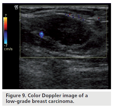
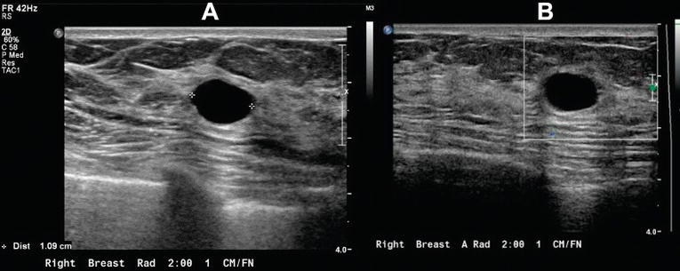
Bildgebung 614 291294 1994. A grayscale image of a palpable breast mass that could be cystic or solid and b color doppler images demonstrate internal vessels thus this is a solid lesion.
Https Onlinelibrary Wiley Com Doi Pdf 10 7863 Jum 1996 15 12 835
Como lo hacemos nosotros.

Breast cancer color doppler ultrasound color meanings. The longitudinal transverse axis ratio and hilar status on color doppler flow and gray scale us images were prospectively studied for each of 145 axillary nodes in 135 women 74. However in the daily clinical practice it has become an indispensable tool and an integral part of the breast ultrasound us. The contribution of color doppler sonography in the study of breast cancer remains a topic of discus sion.
The internal blood flow is consistent with a solid mass such as this patients dermatofibrosarcoma but not with a superficial sebaceous cyst protein containing cyst or hematoma. A small study found that a type of three dimensional 3 d ultrasound picture of the breast can help doctors decide if a breast mass is cancer or not. Diagnosis of the breast tumor entity with vocal fremitus in ultrasound diagnosis.
The age of the patient large size of tumor and typical ultrasound features are highly suggestive of this being a phyllodes tumor of the left breast. Of 1124 breast lesions 590 525 were benign and 534 475 were malignant. To study the flow indices of color doppler ultrasound in the differentiation of malignant and benign breast tumors data of 1124 female patients with breast lesions were analyzed.
A study has shown that using ultrasound in addition to mammography increases the sensitivity to 775 compared to that of mammography alone 50 in women with dense breasts and increased risk of breast cancer. Color doppler sonography can figure 1. The use of color doppler ultrasonography cdus for characterizing breast lesions has increased in recent years.
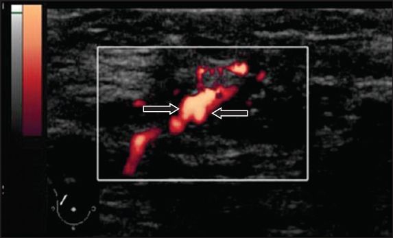
The images above show minimal internal vascularity on color doppler ultrasound and 3 d ultrasound bottom left show the internal lobulation with typical leaf like pattern on sonography. Color flow doppler image displayed in black and white shows blood flow in vessels within the mass arrows. To document differences in color doppler flow and gray scale ultrasonographic us features between benign and malignant axillary lymph nodes in women with primary breast cancer.
The use of breast ultrasound color doppler vascular pattern morphology improves diagnostic sensitivity with minimal change in specificity. 2010 epub ahead of print. Doppler color en el estudio de la mama.
In this study quantitative color doppler radiomics features were algorithmically extracted from breast sonograms for machine learning producing a diagnostic model for breast cancer with higher performance than models based on grayscale and clinical category from the breast imaging reporting and data system for ultrasound bi radsus. Sohn c baudendistel a bastert g. Color doppler of a simple cyst.
The 3 d ultrasound method is called speed weighted 3 d power doppler and it shows blood flow in and around a breast tumor. Color doppler scanning detected vascularity in 505 856 of benign lesions and 509 953 of malignant tumors. If a mass is found in your breast your doctor usually will recommend a.

Breast Ultrasound Leaders In Pharmaceutical Business Intelligence Lpbi Group

Breast Ultrasound Past Present And Future Intechopen

Complex Cystic Breast Masses In Ultrasound Examination Sciencedirect

Color Doppler Sonography Characterizing Breast Lesions
Https Onlinelibrary Wiley Com Doi Pdf 10 7863 Jum 1995 14 2 125
Http E Ultrasonography Org Upload Usg 17043 Pdf

Evaluation Of Solid Breast Lesions With Power Doppler Value Of Penetrating Vessels As A Predictor Of Malignancy Smj