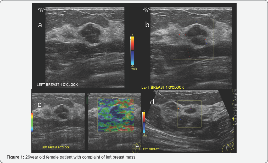
1 they account for 68 of all breast masses and a large proportion of breast biopsies. These findings suggest a diagnosis of giant fibroadenoma of the left breast.

Cancer Therapy And Oncology International Journal Ctoij
In these cases doctors do not recommend having the tumors removed.

Breast cancer fibroadenoma ultrasound image. This breast cancer ultrasound image shows changes related to breast cancer that are not seen as microcalcifications or a mass or lump. When a lesion has the typical features of a fibroadenoma on ultrasound and there are no clinical red flags they can be safely followed clinically. As aforesaid fibroadenoma is a benign condition.
Unlike a breast cancer which grows larger over time and can spread to other organs a fibroadenoma remains in the breast tissue. Theyre pretty small too. 2 fibroadenomas most commonly present as a single painless firm mobile mass but can be.
Fibroadenoma surgery may involve removing a margin of breast tissue surrounding the fibroadenoma. Fibroadenomas often stop growing or even shrink on their own without any treatment. Breast ultrasound uses sound waves to image the tissues of the breast.
An instrument called a transducer sends high frequency sound waves into the breast and captures returning echoes. By measuring echo waves it is possible to determine an objects size shape and consistency. When lesions enlarge or have atypical imaging findings ultrasound guided core biopsy is a minimally invasive outpatient procedure that will give a diagnosis with virtually no complications.
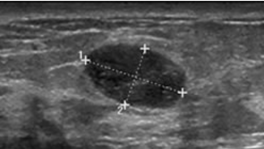
A biopsy taking out breast tissue to check it in the lab is needed to know if a tumor is a fibroadenoma or some other problem. Breast ultrasound showing abnormalities normal breast implants leak breast cancer cysts lactating fibroadenoma papilloma and carcinomas and mondor cord. Old female patient show a large the mass measured 82 cms more or less homogenous well defined mass with posterior acoustic enhancement.
They present most frequently between ages 20 and 50 with peak incidence reported at 20 to 24 years. Fibroadenoma is the most common benign breast tumor in women younger than age 30. Some fibroadenomas are only found on an imaging test like a mammogram or ultrasound.
Rather the right breast is seen as smaller than the left breast. You can also see some benign calcifications scattered in. There is no reported case of fibroadenoma breast cancer.
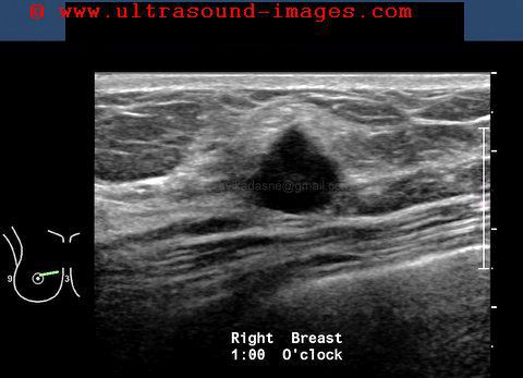
Image shows circumscribed slightly lobulated fibroadenoma. There is no risk of cancer from benign fibroadenoma breast growths. There is a slight increase in the density in the right breast compared with the left.
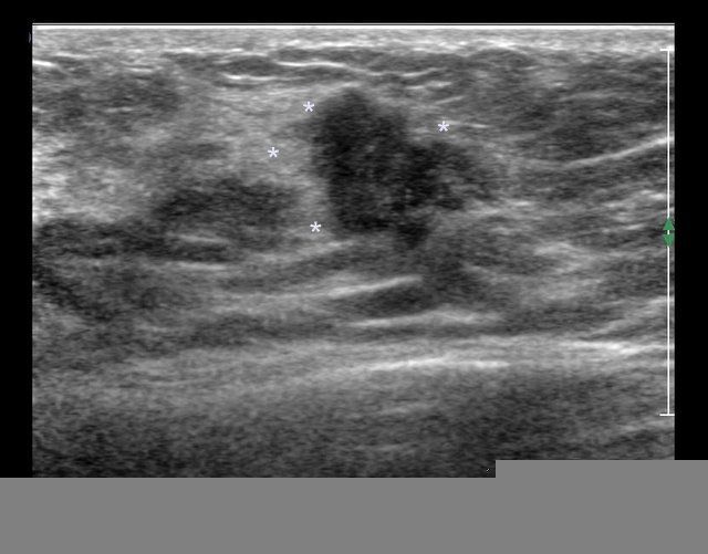
If lumps are not removed they should be periodically checked with the help of imaging tests and physical exams to detect any increase in size. These ultrasound images of the left breast in a 15 yr. Most are only 1 or 2 centimeters in size.

How I Dissolved My Fibroadenoma Without Surgery Predominantly Paleo

Breast And Axilla 5 3 Pitfalls Case 5 3 1 Malignant Lesions Mimicking A Benign Lesion Ultrasound Cases

Ultrasound Can Accurately Diagnose Fibroadenomas In Women Less Than 35 Years
Https Encrypted Tbn0 Gstatic Com Images Q Tbn 3aand9gcsqtreeppb7um7issmxddrcpe7w1m7ug7 Yjfrry9ovbfnlqtzn Usqp Cau

A Gallery Of High Resolution Ultrasound Color Doppler 3d Images Breast

A Gallery Of High Resolution Ultrasound Color Doppler 3d Images Breast
